Fig. 1.

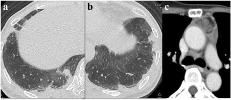
Chest computed tomography scans on admission. a, b Irregular shaped peripheral consolidations in both the lower lobes. c A heterogeneously enhanced mediastinal mass

Chest computed tomography scans on admission. a, b Irregular shaped peripheral consolidations in both the lower lobes. c A heterogeneously enhanced mediastinal mass