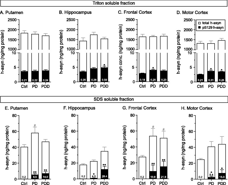
Fig. 9.

Assessment of S129 phosphorylated human alpha-synuclein species in post mortem brain tissue. Frozen human brain tissue samples were received from healthy controls (Ctrl), patients diagnosed with Parkinson’s disease (PD) or Parkinson’s disease with dementia (PDD) (n = 8 per group). Four brain regions were analyzed: putamen (a, e), hippocampus (b, f), frontal cortex (c, g) and motor cortex (d, h). Tissue was sequentially lysed first using 1 % Triton (about 100 ng of protein loaded) (a–d) followed by 1 % SDS containing lysis buffer (about 2 μg of protein loaded) (e–h). Total (open) and pS129 (filled) h-asyn levels are presented in overlapping bars. Numbers inside the bars show the percent of pS129 to total h-asyn. *p <0.05; **p <0.01; #p <0.05; one-way ANOVA followed by a Tukey’s HSD test or Kruskal-Wallis followed by a Dunn’s multiple comparisons test; Error bars indicate SEM
