Fig. 8.

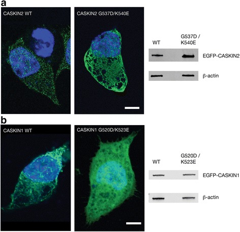
CASKIN2 and CASKIN1 SAM domain expression in Neuro2a cells. a Images were made 48 h after transient transfection with CASKIN2-EGFP (wild type), CASKIN1-EGFP (wild type), mutant CASKIN2 (G537D/K540E)-EGFP, and CASKIN1 (G520D/K523E)-EGFP plasmids. The green fluorescence demonstrates distinct protein distributions for the wild type and mutant proteins. Counterstaining with DAPI (blue) reveals that the subcellular distribution of wild type and mutant proteins in the cytoplasm and nucleus is indistinguishable. Scale bar: 5 μm. b Western blot of cell lysates demonstrating expression of EGFP-CASKIN2 and EGFP-CASKIN1 proteins probed with monoclonal anti-EGFP antibodies. The blot was reprobed with monoclonal anti-β-actin antibodies as a loading control
