
Fig. 5.
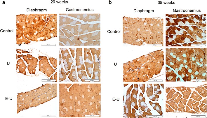
a Representative examples of stained muscle fibers in diaphragm and gastrocnemius muscles of control (top panel), U group (middle panel) and E–U group (bottom panel) in the 20-week cohort of animals. Myofibers positively stained with the anti-MyHC type II antibody appear in brown color (calibration bar 200 μm). Type I fibers appear in white color (not stained). b Representative examples of stained muscle fibers in diaphragm and gastrocnemius muscle of control (top panel) and U group (middle panel) and E–U group (bottom panel) in the 35-week cohort of animals. Myofibers positively stained with the anti-MyHC type II antibody appear in brown color (calibration bar 200 μm). Type I fibers appear in white color (not stained). anti-MyHC anti-myosin heavy chain, E–U elastase–urethane, U urethane