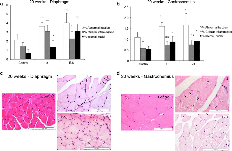
Fig. 7.

a Mean values and standard deviation of the percentage of abnormal fraction (white bars), cellular inflammation (grey bars) and internal nuclei (black bars) in the diaphragm muscle of the 20-week cohort animals. Statistical significance is represented as follows: *p ≤ 0.05 and ***p ≤ 0.001 between any of the intervention groups (U and E–U) and control mice. b Mean values and standard deviation of the percentage of abnormal fraction (white bars), cellular inflammation (grey bars) and internal nuclei (black bars) in the gastrocnemius muscle of 20-week cohort animals. Statistical significance is represented as follows: n.s.: non-significant and *p ≤ 0.05 between any of the intervention groups (U and E–U) and control mice. c Representative examples of muscle structural abnormalities in diaphragm of 20-week cohort animals (calibration bar 100 μm). A representative image of muscles in U group (right top image), in which black arrows point towards inflammatory cells, and a representative image of muscles in E–U group (right bottom image), in which black arrows point towards inflammatory cells and internal nuclei. d Representative examples of muscle structural abnormalities in gastrocnemius of 20-week cohort animals (calibration bar 100 μm). A representative image of muscles in U group (right top image), in which black arrows point towards inflammatory cells, and a representative image of E–U group (right bottom image), in which black arrows point towards internal nuclei. E–U elastase–urethane, U urethane
