Fig. 2.

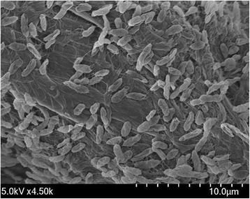
Scanning electron micrograph of Pseudomonas lutea OK2T. The image was taken under a Field Emission Scanning Electron Microscope (FE-SEM, SU8220; Hitachi, Japan) at an operating voltage of 5.0 kV. The scale bar represents 10.0 μm

Scanning electron micrograph of Pseudomonas lutea OK2T. The image was taken under a Field Emission Scanning Electron Microscope (FE-SEM, SU8220; Hitachi, Japan) at an operating voltage of 5.0 kV. The scale bar represents 10.0 μm