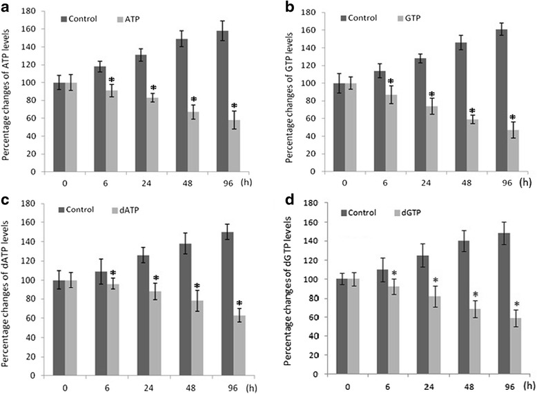
Fig. 3.

Histograms of time-dependent alterations of ATP, GTP, dATP and dGTP levels in embryonic brain tissue on gestation day 11.5. At the indicated time after dosing (40 mg/kg body weight DDATHF), the samples (3–4 embryonic brain tissues as one sample) were collected and handled as described in Methods. The black bars show are the control groups, the grey bars show the ATP, GTP, dATP, dGTP levels respectively of the DDATHF injected mice *P < 0.05
