Fig. 1.

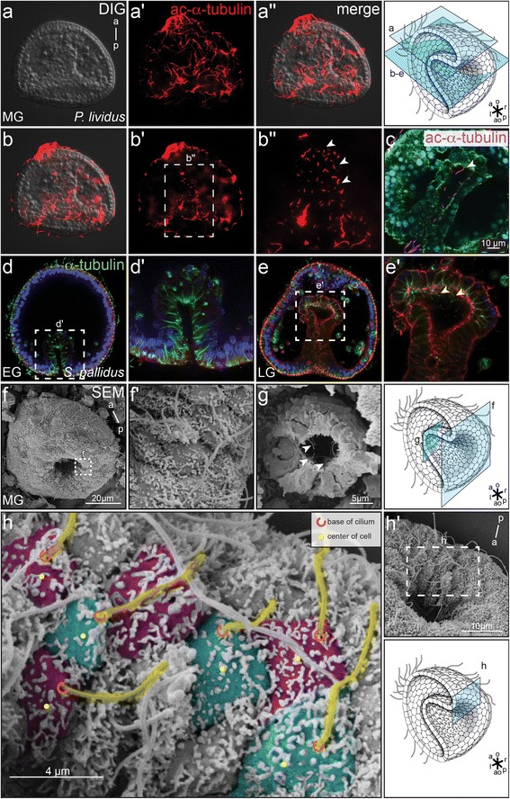
Polarized cilia at the sea urchin archenteron. a–e P. lividus (a–c) and S. pallidus embryos (d–e’) were analyzed by IF for the presence of cilia at the archenteron. Optical sections showed ectodermal (a’; a”) and archenteron cilia (b’–c) at mid gastrula stages. Late (e; e’) but not early S. pallidus gastrula stage embryos revealed cilia in the archenteron and at the archenteron tip (d, d’). Cilia were stained with an antibody against acetylated-α-tubulin (red, a’–c) or anti-α-tubulin (green, d–e’), nuclei were stained with DAPI (d–e’), and cell boundaries were visualized by phalloidin-green (c) or phalloidin-red (d–e’). f, g SEM analysis of P. lividus ectodermal and archenteron cilia. Fractured embryos allowed the visualization of monocilia on archenteron cells (g). Cilia are highlighted by an arrowhead (f–h’) Posterior polarization of cilia on cells which invaginated into the archenteron. Cilia are colored in yellow and individual cells alternating in green and purple. The ciliary base is marked by a red semicircle, the center of the cell is indicated by a yellow dot. Schematic drawings adapted from Blum et al. 2014 [3]
