
Fig. 2.
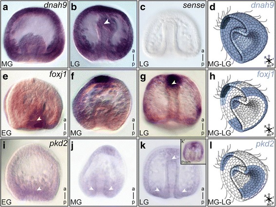
Motile cilia marker genes dnah9 and foxj1 are expressed throughout sea urchin gastrulation. Whole mount in situ hybridization of early (EG), mid (MG) and late (LG) gastrula stage P. lividus embryos, as well as prism stages (k’) for mRNA expression of dnah9 (a–d), foxj1 (e–h) and pkd2 (i–l). Schematic representation of staining in mid-gastrula embryos is highlighted in drawings in (d, h and l). White arrowheads highlight vegetal blastopore and archenteron tip expression areas. Schematic drawings adapted from Blum et al. 2014 [3]