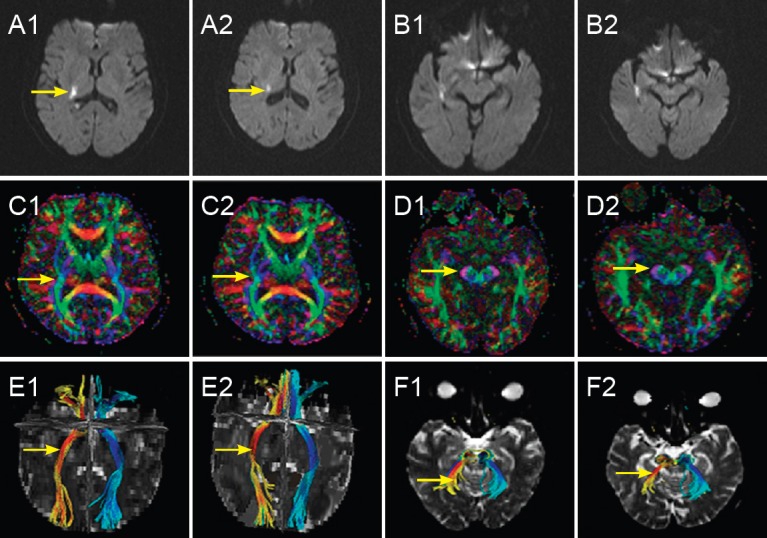
Figure 2.

Alterations in a 77-year-old female patient with acute cerebral infarction in the right basal ganglia in the ozone treatment group before and after treatment.
(1, 2) Pretreatment (1) and 9 days posttreatment (2) in the ozone treatment group, respectively. (A, B) Diffusion weighted images of the infarction focus and corresponding cerebral peduncle before and after treatment: signal intensity was lower in the infarction focus after treatment compared with before treatment (arrows), while no obvious abnormal signal was detected in the corresponding cerebral peduncle. (C, D) Fractional anisotropy images of the infarction focus and corresponding cerebral peduncle before and after treatment: signal intensity in the infarction focus and corresponding cerebral peduncle was lower than in contralateral regions before treatment (arrows), but showed a decreased trend after treatment. (E, F) Three-dimensional images of reconstructed fiber bundles in the infarction focus and corresponding cerebral peduncle before and after treatment: corticospinal tracts (arrows) were injured and aggravated after treatment. Yellow represents longitudinal fiber bundles on the infarction side; green represents longitudinal fiber bundles on the normal contralateral side; red represents horizontal fiber bundles on the infarction side; and blue represents horizontal fiber bundles on the normal contralateral side.
