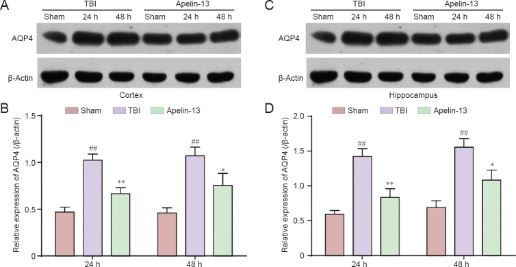
Figure 3.
Apelin-13 acutely reduces AQP4 protein expression in the cortex and hippocampus at 24 and 48 hours post-TBI.
(A, C) Representative western blots of AQP4 protein the cortex (A) and hippocampus (C) were detected by western blot assay. (B, D) Quantitative analysis of AQP4 protein expression in the cortex (B) and hippocampus (D). Optical density of the respective protein bands were analyzed by Quantity One (Bio-Rad) and normalized to β-actin. Data are expressed as the mean ± SEM (6 mice in each group). Statistical comparisons were performed by analysis of variance followed by Dunnett’s t-test. *P < 0.05, **P < 0.01, vs. TBI group; ##P < 0.01, vs. sham group. AQP4: Aquaporin-4; TBI: traumatic brain injury; h: hours.

