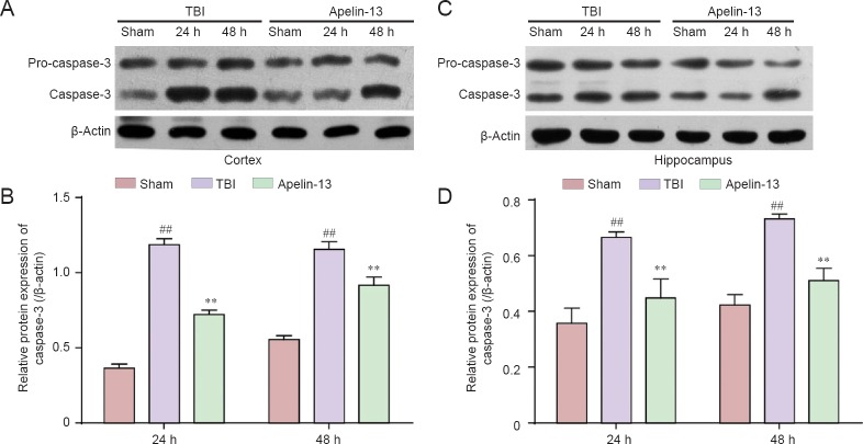
Figure 5.
Inhibition of TBI-induced caspase-3 expression in the cortex and hippocampus by apelin-13 at 24 and 48 hours post TBI.
(A, C) Representative western blots of caspase-3 protein in the cortex (A) and hippocampus (C) were detected by western blot assay. (B, D) Quantitative analysis of caspase-3 protein expresssion in the cortex (B) and hippocampus (D). Data are expressed as the mean ± SEM (6 mice in each group). Statistical comparisons were performed by analysis of variance followed by Dunnett’s t-test. **P < 0.01, vs. TBI group; ##P < 0.01, vs. sham group. TBI: Traumatic brain injury; h: hours.

