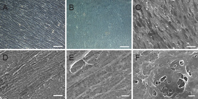
Figure 2.

Morphology of hUCMSC-derived ECM.
(A) Phase-contrast microscopy image and (C) scanning electron microscopy image of hUCMSC-derived ECM before decellularization. (B) Phase-contrast microscopy image and (D) scanning electron microscopy image of hUCMSC-derived ECM after decellularization. (E) Higher magnification of the dotted area in D. (F) Higher magnification of the dotted area in E. Scale bars: A–C, 100 μm; D, 10 μm; E, 2 μm; F, 200 nm. ECM: Extracellular matrix; hUCMSCs: human umbilical cord-derived mesenchymal stem cells.
