Figure 3.
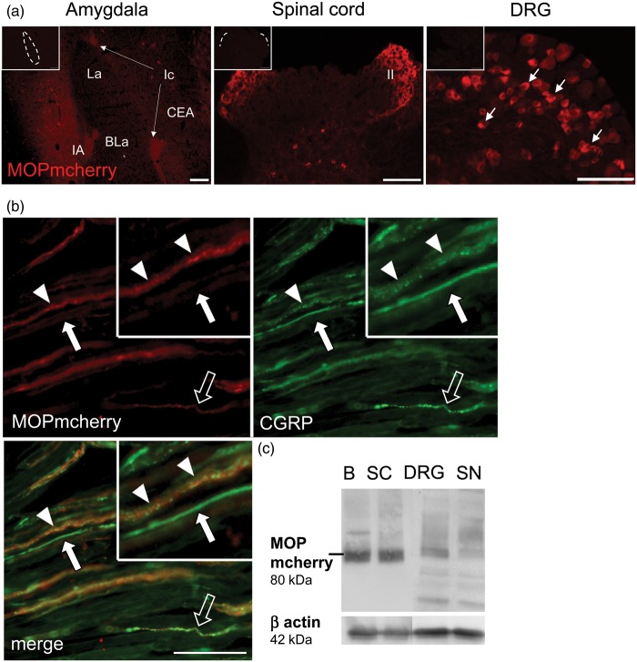
MOP-mcherry immunodetection in knock-in mice. (a) Immunolabeling in cryosections is shown in amygdala (left panel), superficial laminae I/II of spinal dorsal horn (middle panel), and small/medium DRG neurons (right panel). Controls testing for unspecific antibody binding are inserted in the upper left corner (scale bars 100 µm, nuclei of the amygdala: CEA: central nucleus; La: lateral nucleus; BLa: basolateral nucleus; Ic: intercalated nuclei). (b) Longitudinal section of MOP-mcherry knock-in sciatic nerve labeled for colocalization of MOP-mcherry (red) with CGRP (green) in putative nociceptive fibers and fiber bundles (arrowheads: high MOP-mcherry immunoreactivity; open arrow: low MOP-mcherry immunoreactivity; closed arrows: CGRP-ir only, scale bar 20 µm). (c) Western blots on tissues from MOP-mcherry mice confirm MOP-mcherry (80 kDa) localization in brain (B), spinal cord (SC), DRG, and sciatic nerve (SN) relative to β-actin (42 kDa) (all representative images, n = 3).
MOP: µ-opioid receptor; DRG: dorsal root ganglion; CGRP: calcitonin gene-related peptide.

