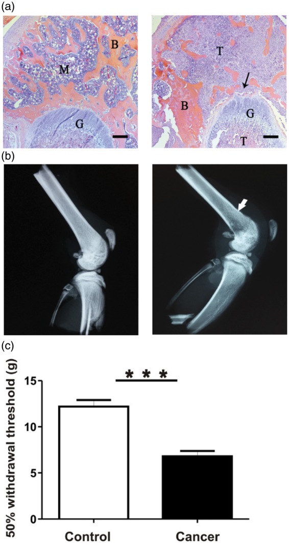
Figure 2.

Model confirmation: structural and histological differences. (a) Representative H&E stained 4 µm thick sections of the ipsilateral distal epiphysis of femurs from control (left) and cancer rats (right). Control bone appears healthy and free of indications of pathology induced by sham injections. In contrast, cancer bone features extensive invasion of cancer cells into areas of the bone marrow and mineralized bone; surfaces of trabecular bone appear ragged and eroded (indicated by arrow). B: mineralized bone; M: marrow; T: tumour cells; G: growth plate. Scale bar represents 300 µm. (b) Representative radiographs of ipsilateral hind limbs of control (left) and cancer rats (right). Control bone appears pathology-free, while cancer bone displays structural modifications and lytic lesions at the injection site in the distal femur epiphysis. All images were taken following fixation of samples from animals 7 to 14 days after model induction. (c) Comparison of 50% withdrawal thresholds between control and cancer groups. Withdrawal threshold to mechanical stimulation of the plantar surface of the ipsilateral hind paw with von Frey filaments was recorded immediately prior to the acute electrophysiological experiment in control (n = 15) and cancer (n = 15) animals. Data are shown as mean ± SEM. ***p < 0.001.
H&E: hematoxylin and eosin;
