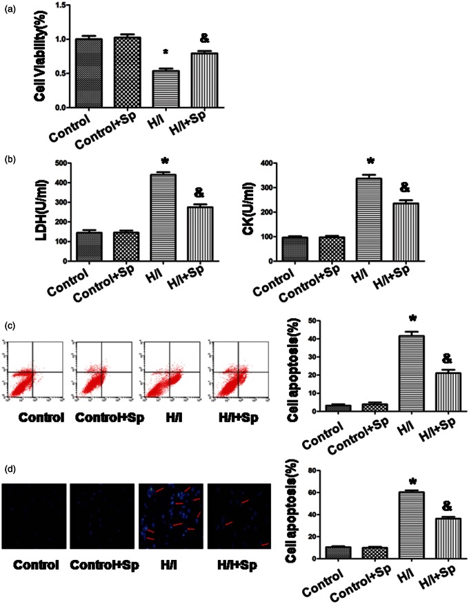
Figure 3.
Spermine inhibited the effects of hypoxia/ischemia on cardiomyocytes apoptosis. (a) Cell viability was measured by CCK-8 assay. The cells that were incubated with the control medium were considered to be 100% viable. (b) The extent of cellular injury was detected by LDH and CK activity. (c) Apoptosis was analyzed by flow cytometry. (d) Detection of nuclear morphology in apoptotic cells using Hoechst 33342 nuclear staining. Apoptotic cells were marked as cells with condensed, disrupted nuclei (arrow, × 400). Apoptotic cells were counted in at least 10 random fields. All the data are mean ± SEM for n = 8 independent experiments. *p < 0.05 versus control group; &p < 0.05 versus H/I group. (A color version of this figure is available in the online journal.)

