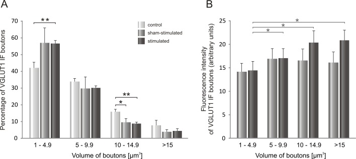
Fig 5.
A. The distribution of VGLUT1 IF boutons of different volume apposing to LG α-motoneurons after seven days of low-threshold stimulation of proprioceptive fibers in the tibial nerve. B. Intensity of VGLUT1 IF signal in boutons apposing to LG α-MNs plotted in function of their volume on the sham-stimulated and electrically-stimulated side. Data are reported as mean +/- SEM. Stimulation of Ia fibers resulted in an increased number of VGLUT1 IF terminals of the smallest size at the expense of the largest (volume > 10 μm3) terminals (*p<0.03, **p<0.005, Mann-Whitney U test). An intensity of the signal tended to increase in the largest boutons.

