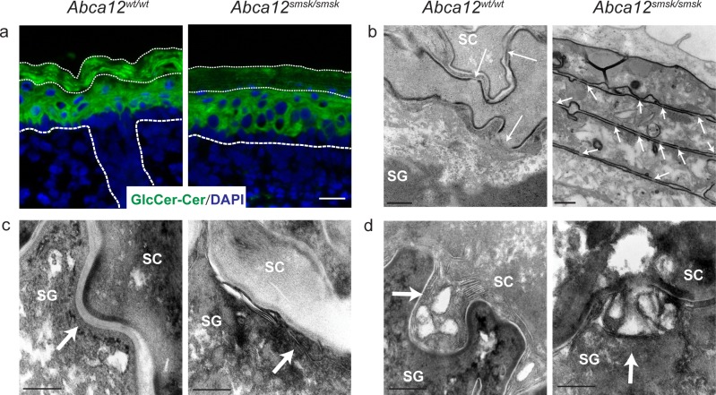
Fig 3. Defects in lipid accumulation in the stratum corneum of Abca12smsk/smsk mutant mice.
(a) Immunofluorescence staining for glucosylceramide/ceramide (GlcCer/Cer) showed localization throughout the suprabasal layers of WT epidermis, whereas reduced levels were detected in the SC (thin dashed lines) of E18.5 Abca12smsk/smsk epidermis. Bar = 25 μm. (b) Transmission electron microscopy (TEM) shows the disappearance of corneodesmosomes (CDs) (white arrows) above the SG-SC interface in WT mice, whereas CDs are retained in smsk SC. Bars = 200 nm and 500 nm respectively. (c) TEM pictures show the presence of normal intercellular lipid lamellae (arrow) at the junctions between SG and SC layers in the WT epidermis but not in the mutant epidermis. Bars = 200 nm. (d) Ultrastructural analysis shows that lamellar bodies (LBs) in WT epidermis were loaded with lipid lamellae and fused with the surface of granular cells (arrow). LBs in mutant epidermis had no lamellar cargo, but fusion with the granular cell membrane appeared normal. Bars = 200 nm.

