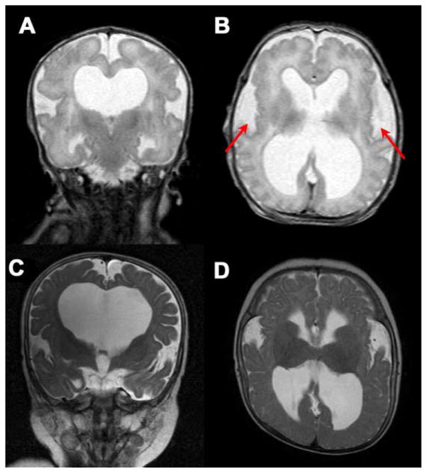
FIG. 2.
T2-weighted MRI images of the brain. A: Coronal view at 3 days of age. Note dilatation of the lateral ventricles and absence of the septum pellucidum. B: Ventriculomegaly, abnormal opercularization, and mildly effaced gyral pattern at age 3 days. C: Progression of hydrocephalus at age 8 months. D: Similar brain abnormalities were still evident at 8 months of age.

