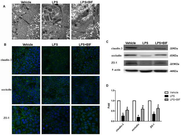
Fig 5. Bifidobacterium prevented the disruption of TJ in vitro.
(A) Electron micrographs reveal the changes of intact TJ in vehicle group, LPS group and LPS+BIF group. (B) Immunofluorescence staining of TJ proteins localization in Caco-2 cells with or without LPS and BIF. Magnification: ×40. (C) Western blot for TJ proteins. Caco-2 cells were grown and treated with LPS and BIF and lysed. The lysates were used for immunoblotting for claudin-3, occludin, ZO-1 and β-actin. Representative results of one experiment are shown. Similar results were obtained in three independent experiments: vehicle group, LPS group, LPS+BIF group. (D) The intensity of the bands was quantified by scanning densitometry, standardized with respect to β-actin protein and expressed as mean ± SD fold change compared with vehicle cells. *P < 0.01 vs the vehicle group, #P < 0.01 vs the LPS group.

