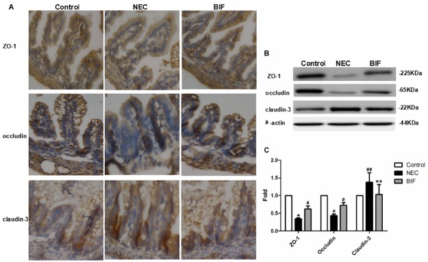
Fig 6. Bifidobacterium prevented the disruption of TJ in a rat NEC model.
(A) TJ proteins localization was evaluated by Immunohistochemical staining in the terminal ileum of neonatal rats. Representative slides for control, NEC, and BIF were shown. Magnification: ×40, n = 3 to 6 per group. (B) Western blot for TJ proteins. Terminal ilea were subjected to immunoblotting for ZO-1, occludin claudin-3 and β-actin. Representative results of one experiment are shown. Similar results were obtained in three independent experiments: control group, NEC group, BIF group. (C) The intensity of the bands was quantified by scanning densitometry, standardized with respect to β-actin protein and expressed as mean ± SD fold change compared with control animals.*P < 0.01, ##P < 0.05 vs the control group, #P < 0.01, **P < 0.05 vs the NEC group.

