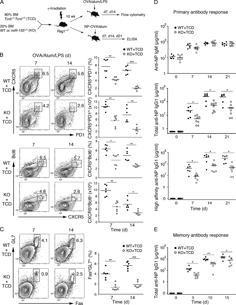
Figure 3.
T cell–intrinsic requirement of miR-155 for generating GC B and Tfh cells. (A–D) Generation and immunization of mixed BM chimera mice with T cell–specific loss of miR-155. Sublethally irradiated Rag1−/− mice were reconstituted with BM cells from miR-155−/− and Tcrb−/−Tcrd−/− (TCD) or WT and TCD mice at a 1:4 ratio and subsequently immunized with OVA/alum/LPS (B and C) or NP-OVA/alum (D). (B, left) Flow cytometry analysis of Tfh cells. Numbers adjacent to the outlined areas indicate percentages of CXCR5hiPD1hi (top) and CXCR5hiBcl-6+ (bottom) Tfh cells among total CD4+ T cells. (Right) Summary of the percentage and cell number of CXCR5hiPD1hi and CXCR5hiBcl-6+ Tfh cells. (C, left) Flow cytometry analysis of GC B cells. Numbers adjacent to the outlined areas indicate the percentage of GC B cells (FashiGL7hi) among total B cells (B220+). (Right) Summary of the percentage of GC B cells. (D) ELISA analysis of NP-specific IgM (top), total NP-specific IgG1 (middle), and high-affinity anti-NP IgG1 (bottom) antibodies in the sera from NP-OVA/alum–immunized mice. (E) Mixed BM chimera mice in Fig. 3 A were reimmunized with NP-OVA 3 mo after primary immunization. Total anti-NP IgG1 in the sera was determined by ELISA. Each dot represents an individual mouse. Horizontal lines indicate the mean. *, P < 0.05; **, P < 0.01; ***, P < 0.001 (Student’s t test). Data were combined from two independent experiments (B and C). n = 6–8.

