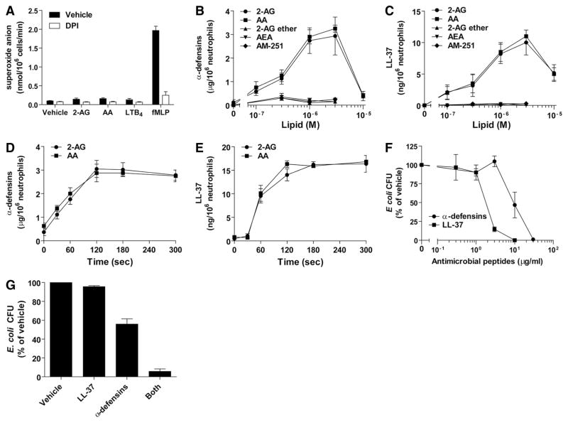
Figure 3. 2-AG and AA induce the release of antimicrobial factors.
(A) Human neutrophils (2×10⁷ cells/ml) in HBSS containing 1.6 mM CaCl2 were incubated with 1 μM cytochalasin B for 15 min and then stimulated with 3 μM 2-AG, 3 μM AA, 100 nM LTB4, or 100 nM fMLP for 5 min in the presence or absence of 10 μM DPI. Incubation was stopped by placing the samples in an ice-water bath and analyzed for O2− production, as described in Materials and Methods. (B and C) Human neutrophils (2×10⁷ cells/ml) in HBSS containing 1.6 mM CaCl2 were stimulated with 2-AG, AA, 2-AG ether, AEA, or AM-251 at the indicated concentration for 5 min. Incubations were stopped by placing the samples in an ice-water bath, and then cell-free supernatants were analyzed for their content in α-defensins or LL-37, as described in Materials and Methods. (D and E) Human neutrophils (2×10⁷ cells/ml) in HBSS containing 1.6 mM CaCl2 were stimulated with 3 μM 2-AG or AA for the indicated times. Incubations were stopped by placing the samples in an ice-water bath, and then the cell-free supernatants were analyzed for their content in α-defensins or LL-37, as described in Materials and Methods. (F) Purified LL-37 or α-defensins were incubated with exponentially growing E. coli suspensions (10,000 CFU/ml) in sodium phosphate buffer containing 1% TSB for 4 h at 37°C. (G) Purified LL-37 (40 ng/ml), α-defensins (10 μg/ml), or both were incubated with exponentially growing E. coli suspensions (10,000 CFU/ml) in sodium phosphate buffer containing 1% TSB for 4 h at 37°C. (F and G) Samples were diluted 1/300 in incubation medium, plated on LB agar plates, and incubated overnight at 37°C to allow CFU enumeration. (A–G) The data represent the mean (±SEM) of at least three independent experiments, each performed in duplicate.

