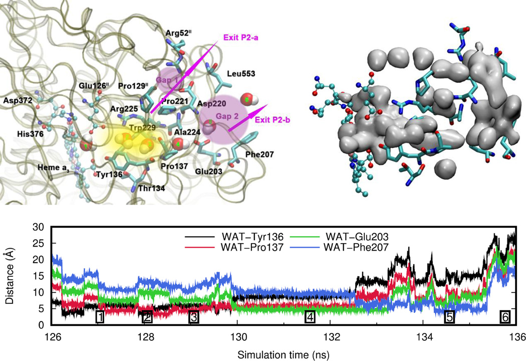
Figure 8.
Water exit pathway P2. Top left: Detailed water pathway highlighting relevant residues. The pathway connects the water pool above the DNC to the outer side of the membrane via a water pocket (yellow) and two regions (pink, Gap1 and Gap2) that open towards the bulk solvent. Gap 1 is surrounded by residues Arg52II, Pro129II, Pro221, Asp220 and Leu553; Gap 2 is surrounded by residues Leu553, Asp220, Ala224, Glu203 and Phe207. Top right: Water occupancy averaged over the entire trajectory (isosurface plot at 25% occupancy), showing the path that connects the water pool to the protein exterior. Bottom: Distances between the center of mass of the water molecule leaving the water pool (WAT) and relevant residues along pathway P2 (Simulation F, see Table 1). The numbers refer to the position of the water molecule as depicted on the left.

