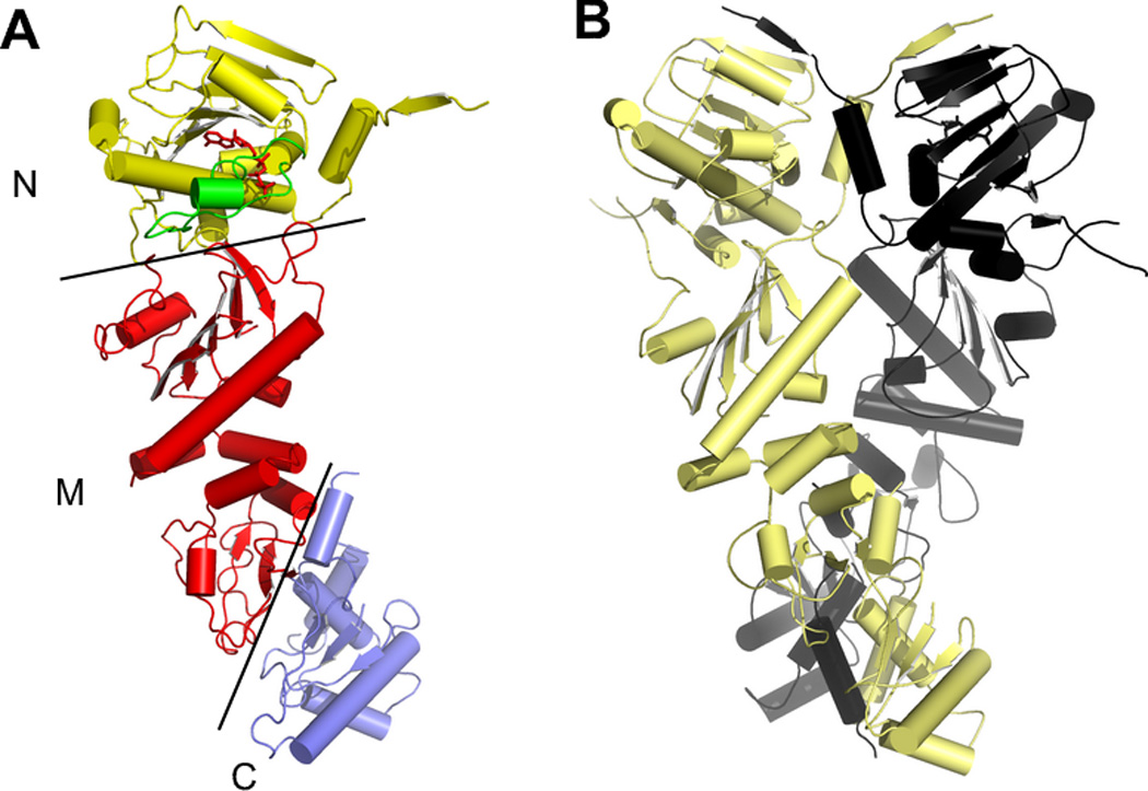
Figure 1.

The structure of full-length yeast Hsp90 (PDB 2cg9). A) One subunit of the dimer, showing the N-terminal, Middle, and C-terminal domains. B) The structure of the intact Hsp90 dimer, showing the dimerization of both the N and C domains. The co-chaperone p23, which was crystallized with the Hsp90, has been omitted for clarity.
