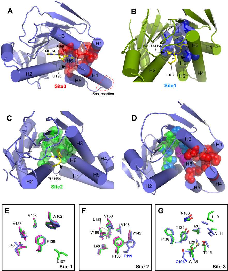
Figure 6.

N-terminal domain structures showing ligand binding sites and overlay of binding site residues from individual paralogs. A) Grp94 in complex with NECA (PDB 1qy5). Site 3 residues are colored red, and the location of the Grp94 5 amino acid insertion is shown in the red box. B) Hsp90α in complex with PU-H54 (PDB 3o0i). Site 1 residues are colored blue. C) Grp94 in complex with PU-H54 (PDB 3o2f). Site 2 residues are colored green. D) Composite of Sites 1, 2, and 3 on Grp94 (PDB 1qy5). Phe199, which is common to all 3 sites is colored magenta. E) Overlay of Site 1 residues from hHsp90:PU-H54 (PDB 3o0i, green), Grp94:SNX0723 (PDB 4nh9, blue), and Trap-1:PU-H71 (PDB 4z1f, magenta). Numbering is for hHsp90; the equivalent of L107 is disordered in the Grp94 and Trap-1 structures. F) Overlay of Site 2 residues from hHsp90:PU-H54 (PDB 3o0i), Grp94:PU-H54 (PDB 3o2f), and Trap-1:PU-H71 (PDB 4z1f). Coloring is as in (E). Numbering is for hHsp90 except for F199 which is from Grp94. G) Overlay of Site 3 residues from hHsp90:apo (PDB 1yes, green) and Grp94:NECA (PDB 1qy5, blue). Trap-1 is not shown due to lack of a suitable model for comparison. Numbering is for hHsp90 except for G196 which is from Grp94.
