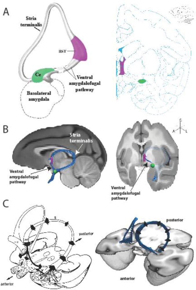
Figure 1.
The EAc and its white matter pathways. A, (left) a schematic of the extended amygdala concept as proposed by Heimer and colleagues, and (right) a plate from a rhesus monkey brain atlas (Paxinos et al. 2009) highlighting the location of the BST (purple) and Ce (green). B, (left) mid-sagittal and (right) rotated coronal MRI slices through a rhesus monkey brain template with an overlaid rendering of the deterministic tractography results showing the pathways connecting the BST and Ce. Also depicted are the waypoint ROIs used in the analysis to define the BST (purple) and Ce (green). Note that the ST in the sagittal view is projected in 3D out in front of the MRI slice. The ST is occluded in the tilted coronal view, traveling caudally behind the MRI slice then arching dorsally and rostrally back out in front of the MRI slice, into the BST waypoint. C, (left) a classic drawing (Roberts 1992) of the major axonal pathways leaving the amygdala, and (right) a 3D tractography rendering of bilateral ST/VA pathways.

