
Figure 5.
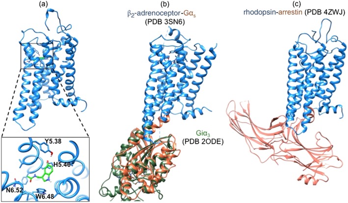
Structures useful for the study of MT1 receptor structure–function relationships. MT1*‐MLT (A) was derived from active forms of rhodopsin, β2‐adrenoceptor and A2A adenosine receptors (unpublished data, N.R.). Docking of melatonin (MLT) in the solvent‐accessible cavity was achieved by energy relaxation by 300‐ns molecular dynamics simulations. The structure of the MT1*‐MLT‐Giα3 complex could be modelled on the basis of the sequence homology with the β2‐adrenoceptor‐Gαs structure (Rasmussen et al., 2011b) and the homology between Gαs and Giα3 (Soundararajan et al., 2008) (B) whereas the structure of the MT1*‐MLT‐arrestin complex could be modelled based on the crystallized rhodopsin‐arrestin complex (Kang et al., 2015) (C).