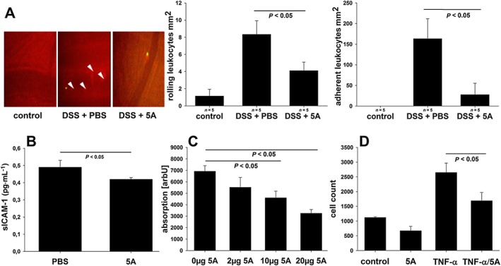
Figure 4.

Effect of 5A peptide on leukocyte–endothelium interactions. (A) C57BL/6 mice on DSS (3% w v‐1) were administered, daily, 5A peptide (0.25 mg·mL−1) or PBS by i.p. injection (n = 5). Leukocyte–endothelium interactions were assessed at day 5 using intravital microscopy as described in Methods. Left panel: original snapshots of peritoneal arterioles. Arrowheads indicate fluorescently labelled adherent leukocytes. Bar graphs: number of rolling (middle panel) and firmly adherent (right panel) leukocytes in postcapillary venules of non‐colitic mice and colitic mice treated with PBS or 5A peptide. (B) Levels of soluble adhesion protein ICAM‐1 were determined in plasma from 5A peptide‐ or PBS‐treated mice by ELISA (n = 5). (C) Effect of 5A peptide on adhesion of BCECF‐AM‐labelled U937 monocytes to HIMECs stimulated with TNFα (10 μg·mL−1) was assessed in vitro using a static adhesion assay. The results from two independent experiments are shown, each in pentuplicate. (D) Effect of 5A peptide on transendothelial migration of calcein‐AM‐labelled U937 monocytes across confluent monolayers of HIMEC grown on polycarbonate pore filters and stimulated with TNFα (10 μg·mL−1). The results from two independent experiments are shown, each in triplicate. Data were analysed using one‐way ANOVA followed by Student–Neuman–Keul of Mann–Whitney test.
