
Figure 10.
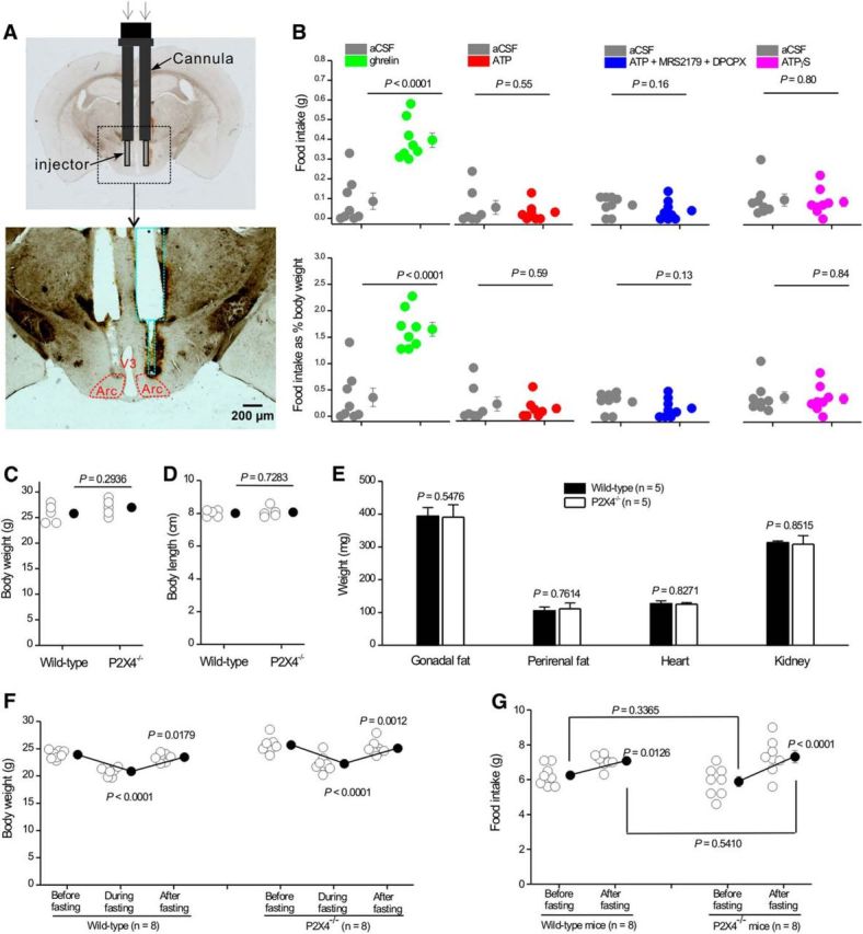
Assessment of feeding in response to ATP and in P2X4 knock-out mice. A, Schematic illustrating the location of the guide cannula and injector for intra-Arc ATP and ghrelin injections. Top, Bilateral guide cannula and injectors projecting out of the cannula implanted in the mouse brain. Bottom, Track of the bilateral cannula with injectors, as marked by green dotted lines in relation to Arc (red). B, Food intake (top graphs) or food intake as a percentage of body weight (bottom graphs) during a 1 h period after bilateral delivery of 500 nl ghrelin (0.7 mm), ATP (3 mm), ATP (3 mm) plus MRS2179 (1 mm), and DPCPX (1 mm) or ATPγS (3 mm). C, D, Body weight (C) and length (D) of 2-month-old wild-type and P2X4 knock-out mice. E, Plot of gonadal fat, perirenal fat, heart, and kidney weights from 2-month-old wild-type and P2X4 knock-out mice. F, Body weights of wild-type and P2X4 knock-out mice before, during, and after fasting. G, Plot of food intake for wild-type and P2X4 knock-out mice before and after fasting.