Figure 1.
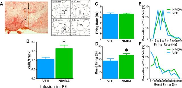
Activation of the RE produced an enhancement of VTA DA neuron population activity. A, Left, Representative image of electrode tracks (solid black arrows) and electrode tip (dashed arrow) in the VTA. Right, Representation of histological placements of infusion cannulae into the RE (open circles). B, Activating the RE with NMDA enhanced the number of spontaneously active DA neurons firing in the VTA (expressed as cells/track, green bar) compared with infusion of vehicle (VEH; blue bar). C, D, The average firing rate of spontaneously active DA cells was not affected by infusion of NMDA into the RE, but the percentage of cells firing in bursts was increased. E, F, Distribution of firing rate and burst firing were not affected by infusion of NMDA into the RE (Kolmogorov–Smirnov test). *p < 0.05 (unpaired t test). VEH, n = 8 rats; NMDA, n = 8 rats; VEH, n = 59 neurons; NMDA, n = 111 neurons. Data are represented as mean ± SEM.

