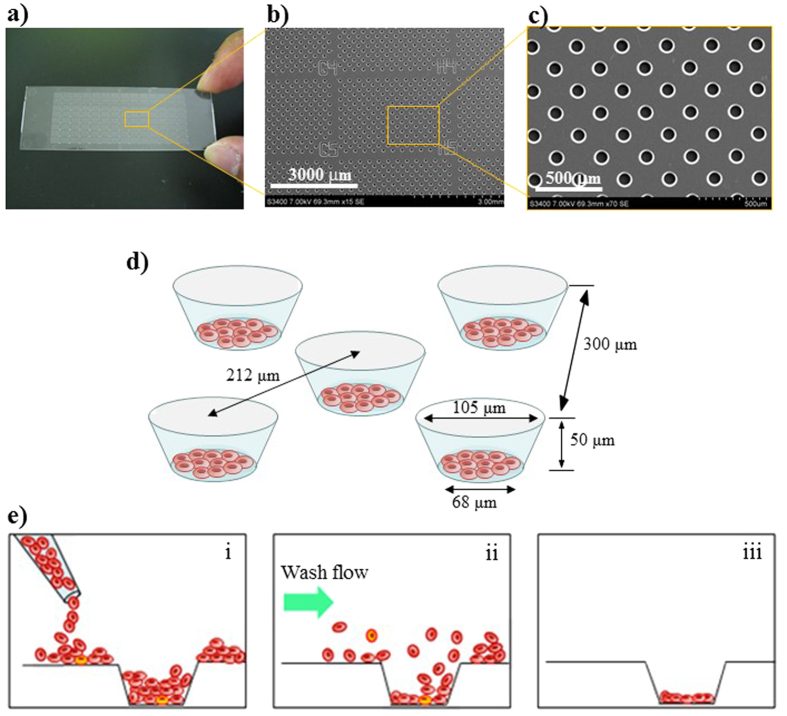
Figure 6. Construction of the cell microarray chip and monolayer formation of erythrocytes in the microchamber.
(a) Photographic and (b,c) SEM images of a cell microarray chip. The cell microarray chip comprised 20,944 microchambers in a plastic slide of glass-slide size. The cell microarray chip had 112 (14 × 8) clusters of 187 microchambers. (d) Each microchamber was 105 μm in width, 50 μm in depth, and 300 μm in pitch, and comprised a frustum with a 68-μm diameter flat bottom for the accommodation of erythrocytes as a monolayer. These images were cited from doi: 10.1371/journal.pone.0013179.g002. (e) Schematic cross-section images of erythrocytes in microchamber, showing (i) dispersing, (ii) washing for removing the excess erythrocytes, and (iii) monolayer formation.

