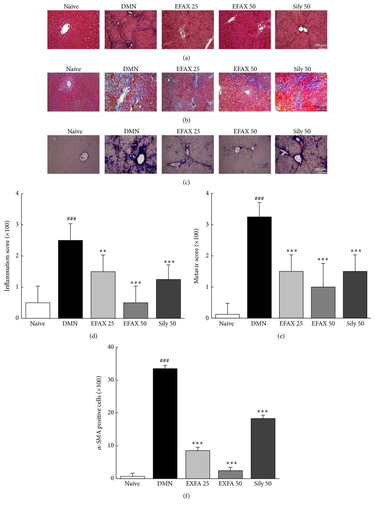
Figure 4.
Histopathological findings and immunohistochemical staining of liver tissues. (a) Hematoxylin and eosin staining (H&E), (b) Masson's trichrome staining, and (c) immunohistochemistry for α-SMA; the histological examinations were performed under light microscopy (×100). (d) The inflammation scores, (e) METAVIR scores, and (f) the number of α-SMA positive cells were analyzed. The data are expressed as the mean ± SD (n = 6). ### p < 0.001 compared with the naive group; ∗∗ p < 0.01, ∗∗∗ p < 0.001 compared with the DMN group.

