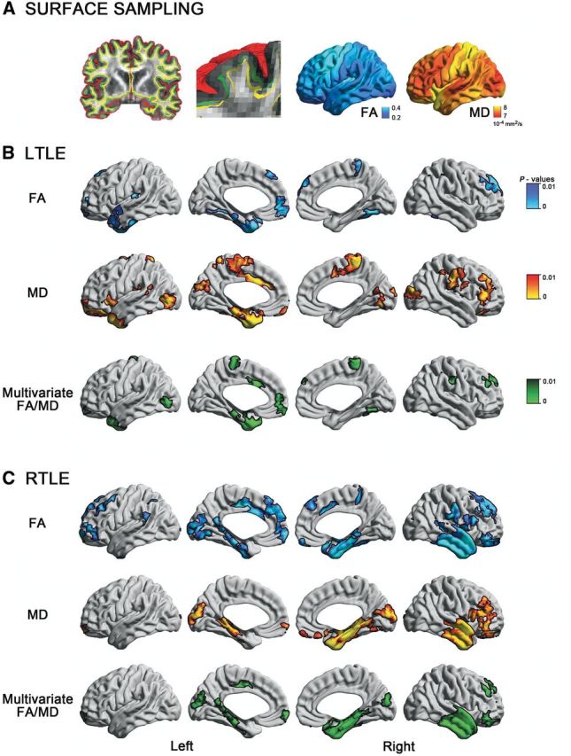
Figure 1.

Surface-based mapping of SWM diffusion in TLE. (A) After generating the inner (white matter–grey matter, green) and outer (grey matter–CSF, red) cortical surfaces, we computed a Laplacian potential field between the white matter–grey matter interface and the ventricular walls to guide placement of a surface running 2 mm below the white matter–grey matter boundary (SWM, yellow). Fractional anisotropy (FA) and mean diffusivity (MD) were sampled on this surface. (B) and (C) show uni- and multivariate SWM diffusion anomalies in left (left TLE) and right (right TLE) patients relative to controls. To correct for multiple comparisons, findings were thresholded at PFWE < 0.05, using random field theory for non-isotropic images (cluster threshold P < 0.01).
