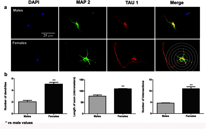
Figure 2. Morphological analysis of neurites in hippocampal neurons from male and female cultures under basal conditions.
(a) Representative examples of male and female neurons on Stage III at 2 DIV. In the right bottom panel, the grid used for Sholl analysis is shown. Neurons are immunostained for microtubule associated protein-2 (MAP2) (green), TAU (red) and DAPI (blue). (b) Number of primary neurites, length of the axon and complexity of the dendritic arbour (number of intersections of dendrites with the circles in the Sholl analysis). Data are the mean ± SEM of 3 hippocampal cultures per sex. **p < 0.01 vs male values.

