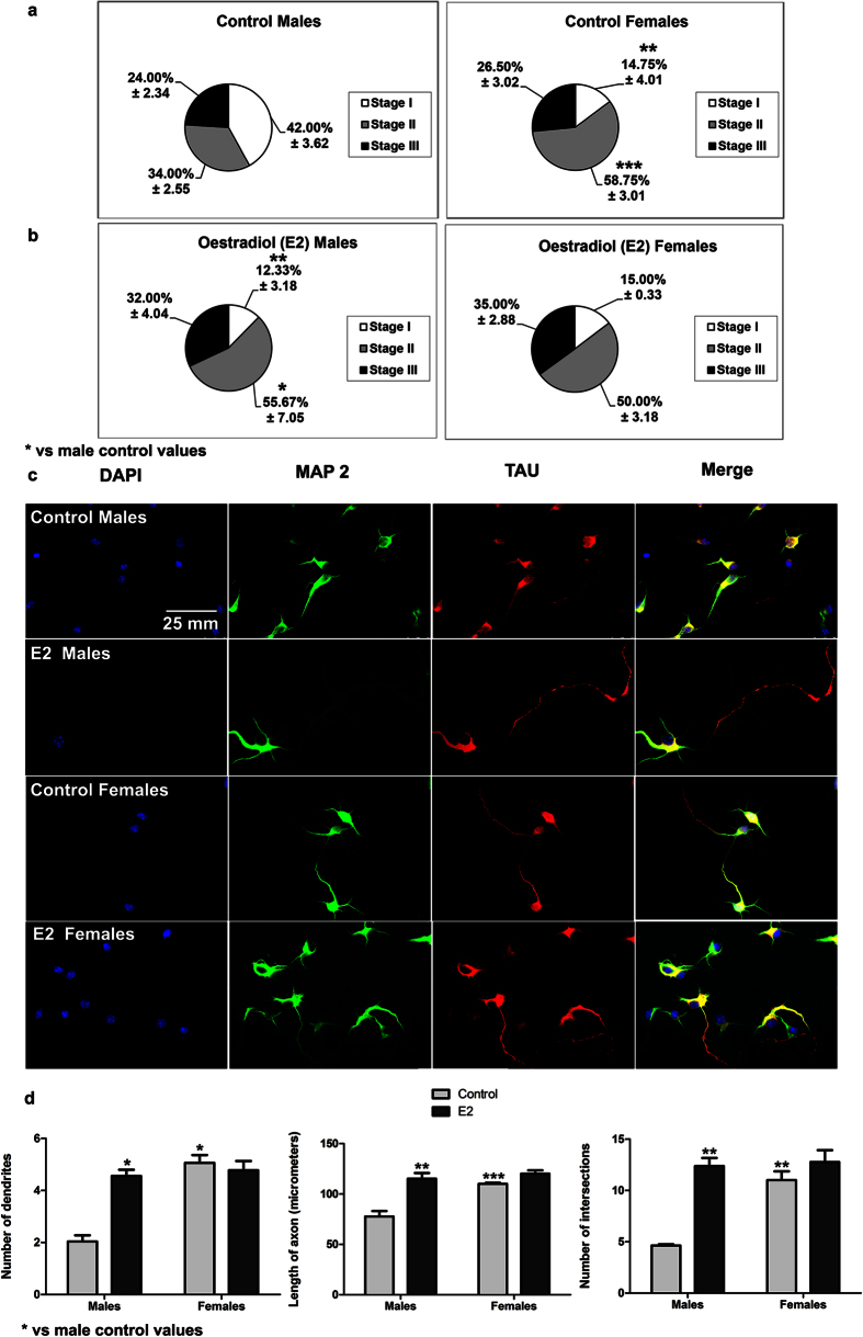
Figure 9. Effect of oestradiol (E2) on neuritogenesis of male and female hippocampal cultures.
(a,b) Proportion of neurons in the three stages of differentiation in male and female cultures at 2 DIV treated 24 h with E2. (c) Representative examples of male and female neurons treated with E2 and immunostained for MAP 2 (green), TAU protein and DAPI (blue) at 2 DIV. (d) Number of primary neurites, length of the axon and complexity of the dendritic arbour (number of intersections of dendrites with the circles in the Sholl analysis). Data are the mean ± SEM of 3 hippocampal cultures. ***p < 0.001, **p < 0.01 and *p < 0.05 vs control male values.

