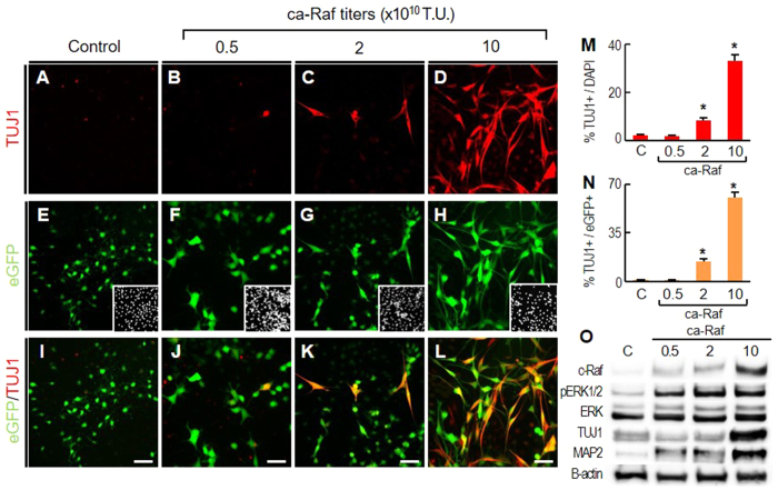
Figure 2. Raf-Erk signaling in NSCs induces neuronal differentiation in a cell-autonomous manner.
NSCs derived from rat embryonic cortices were cultured at low cell density (5–10% cell confluence). These low density cultures were transduced with bicistronic ca-Raf-IRES-eGFP (0.5, 2, and 10 × 1010TU) or control (eGFP, A,E,I). After four days, %TUJ1+ among DAPI+ cells (M) and % TUJ1+ among eGFP+ cells (N) were estimated. *p < 0.001, n = 4, one-way ANOVA. Insets of (E–H), DAPI+ cell images of the same microscopic fields. Erk activation and TUJ1 and MAP2 protein were also measured by WB (O).

