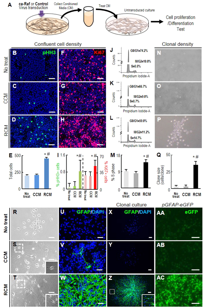
Figure 3. Cortical NSCs transduced with ca-Raf secrete factors that promote cell proliferation and astrocytic maker expression.
(A) Schematic of the experimental procedure. NSCs derived from rat embryonic cortices were cultured at high cell density (80–90% cell confluence) and transduced with ca-Raf or control viruses (10 × 1010TU). Media conditioned by the transduced cultures were collected and non-transduced NSC cultures were exposed to the collected media as described in ‘Materials and Methods’. (B–Q) Cell proliferation estimated from total cell numbers (B–E), %pHH3+ cells (B–D,I), %Ki67+ cells (F–H,I), and % of cells present in S-phase measured by FACS analysis (J–M) grown at normal confluent cell density (50–70% cell confluence). Cell proliferation was further estimated by sizes of clones (cells/clone) formed 4 days after plating NSCs at a clonal density (1000 cells/6-cm dish) (N–Q). Significant different from the untreated* and CCM-treated# at p < 0.001, n = 3 cultures, one-way ANOVA. R-AC, Cell morphology (R–T) and GFAP expression (U-AC). Expression of the astroglial marker GFAP was assessed with an antibody against GFAP in the cultures plated at normal (U–W) and clonal density (X–Z) and by eGFP-filtered microscopic examination in cultures derived from pGfap-eGFP mouse embryonic cortices (AA-AC). Insets, enlarged images of the boxed areas. Scale bars, 100 μm.

