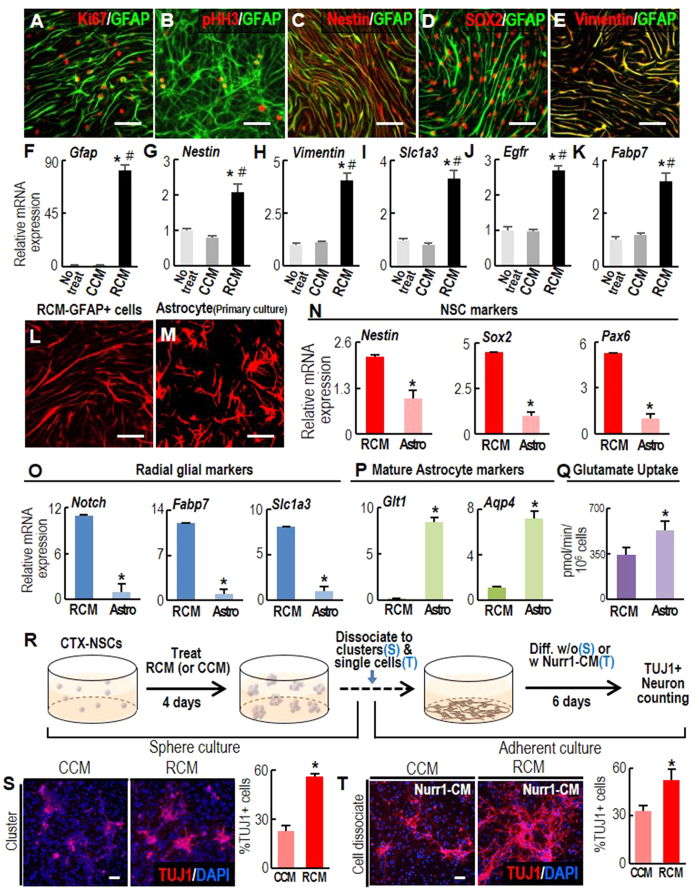
Figure 4. Radial glial- or adult NSC-like properties of the GFAP+ cells generated by Raf-CM treatment.
(A–K) NSC marker expression in the GFAP+ cells generated by RCM treatment (RCM-GFAP+ cells). Immunocytochemical (A–E) and real-time PCR (F–K) analyses were used to compare marker expression in RCM- and CCM-treated cultures. Significant different from the untreated* and CCM-treated# at p < 0.005, n = 3, one-way ANOVA. (L–Q), Morphology (L,M), marker expressions (N–P), and glutamate uptake activity (Q) in RCM-GFAP+ cells (RCM) and primary-cultured astrocytes (Astro). Differentiated astrocytes were cultured from mouse cortex on postnatal day 5. *p < 0.005, n = 3–5, Student’s t-test. (R–V), Neurogenic potential of RCM-GFAP+ cells. (R), Schematic of the experimental procedure. RCM- and CCM-treated neurospheres were dissociated into small clusters (S) or single cell dissociates (T), and plated at identical cell densities in N2 media. Neuronal differentiation was examined without (S) or with Nurr1-CM treatment (T). After six days, neuron yields were assessed as % TUJ1+ cells among total DAPI+ cells. *p < 0.001. n = 3–6 cultures, Student’s t-test. Scale bars, 50 μm.

