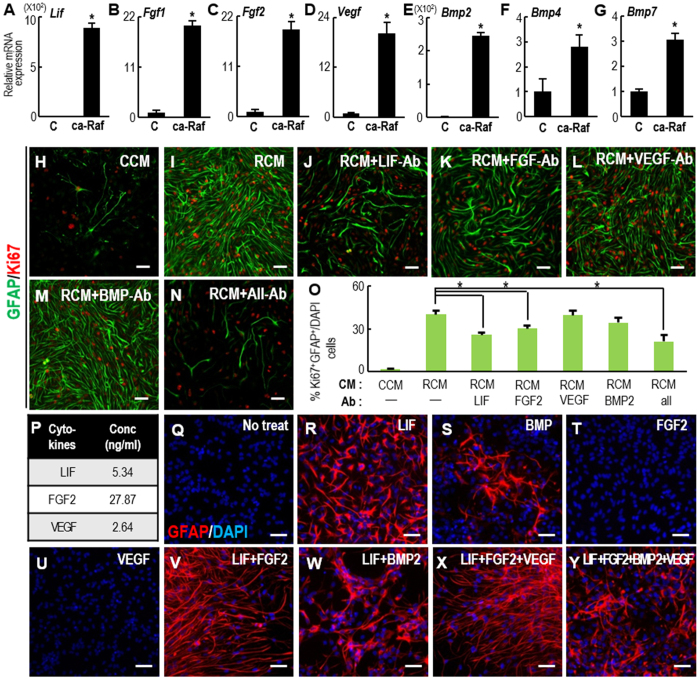
Figure 7. Molecules responsible for RCM induction of proliferating astrocytes.
(A–G) Real-time PCR analyses of candidate molecules known to induce NSC proliferation and astrocytic differentiation. mRNA expression was compared between NSCs transduced with the control and ca-Raf virus. *Significance at p < 0.01, n = 3–6 reactions, Student t-test. (H–O) Treatment with blocking antibody to test its effect on RCM induction of proliferating astrocytes (GFAP+, Ki67+ cells). E14 cortical NSCs were treated with CCM (H) or RCM in the absence (I) or presence (J–N) of the blocking antibodies for 4 days. The antibodies used were anti- LIF, FGF2, VEGF, and BMP (0.1 mg/ml for all above ab). *Significantly different from RCM-treated cultures at p < 0.01, one-way ANOVA, n = 3–5 cultures. (P) Cytokines in the RCM determined with a Bio-plex® 200 System. (Q–Y) Morphology of GFAP+ cells induced by treatment with the indicated cytokines. Cortical NSC cultures were treated with the concentrations of LIF, FGF2, and VEGF determined in (P), and 10 ng/ml of BMP2. Scale bars, 50 μm.

