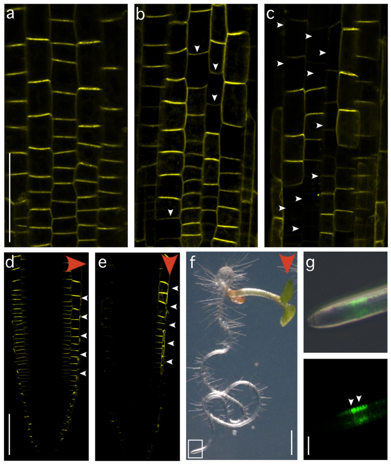
Figure 3. Analysis of a predicted PPP1 DNA-binding site in planta.
(a) PIN2p::PIN2:VENUS expression in eir1-4 root meristem epidermis cells at 4 DAG. (b,c) PIN2pm::PIN2:VENUS signals in root meristems in the progeny of two transformed lines at 4 DAG: Limited (b) and pronounced (c) alterations in reporter expression are indicated by arrowheads. (d) PIN2p::PIN2:VENUS expression gradient after 90 minutes of gravistimulation in a primary root meristem at 4 DAG (red arrowhead: direction of gravity vector; white arrowheads indicate differential PIN2-VENUS abundance). (e) Lateral PIN2pm::PIN2:VENUS expression gradient in a vertically oriented seedling at 4 DAG (red arrowhead: direction of gravity vector; white arrowheads indicate differential PIN2-VENUS abundance). (f) eir1-4 PIN2pm::PIN2:VENUS seedling at 7 DAG grown on a vertically oriented nutrient plate (red arrowhead: direction of gravity vector). (g) Higher magnification of the seedling’s root tip depicted in F (white rectangle). White arrowheads indicate VENUS signal gradient. Bars: a–e = 50 μm; f = 1 mm; g = 75 μm.

