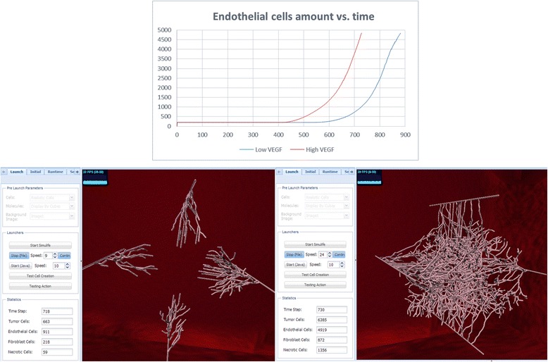
Fig. 11.

High vs. low VEGF secretion simulations. Top: graph of endothelial cells showing that at low VEGF the endothelial cells are activated at ~600ts and reach ~5000 cells at ~900ts, much later than at high VEGF secretion, which begins at ~400ts and reaches ~5000 cells at ~700ts. Bottom: images of these runs in SimuLife (left for low VEGF, right for high VEGF), presenting amounts on their left tab. Both images are presented at approximately the same time step–at low VEGF angiogenesis has only begun, whereas at high VEGF there are many activated and branched vessels
