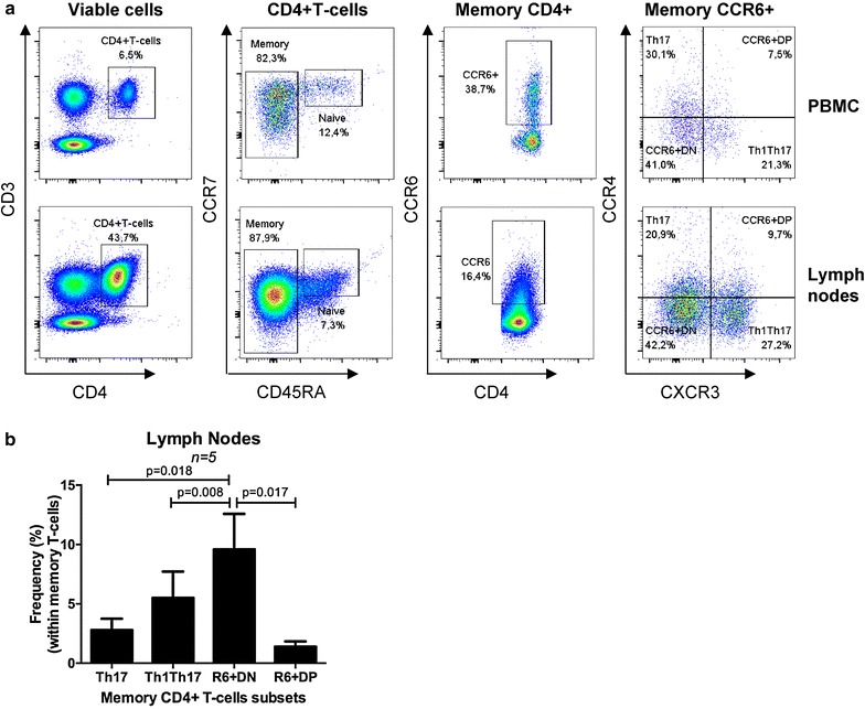
Fig. 7.

CCR6+DN are predominant in lymph nodes of HIV-infected individuals receiving ART. a, b Matched PBMCs and inguinal lymph node cells from three CI on ART individuals (CI 36, CI 37, CI 38; Additional file 8: S2 Table) were stained with a cocktail of fluorochrome-conjugated CD3, CD4, CD45RA, CCR4, CXCR3, CCR6, and CCR7 Abs. A viability staining was used to exclude dead cells. Viable memory CD4+ T-cells (CD3+CD4+CD45RA−) expressing CCR6 were analyzed for their differential expression of CCR4 and CXCR3. The four CCR6+ subsets including Th17, CCR6+DP, CCR6+DN, and Th1Th17 were identified in both PBMCs and cells from lymph nodes. a Shown is the phenotype of PBMCs (upper panels) and lymph node cells (lower panels) in one representative donor. b Shown are statistical analysis of the frequency of CCR6+ subsets in the lymph node (n = 3). Paired t-Test p-values are indicated on the figures
