Fig. 9.

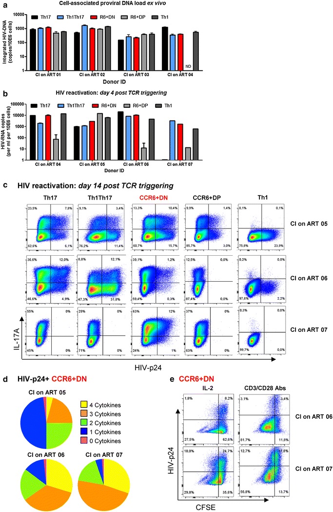
CCR6+DN carry replication competent HIV-DNA. a, b The four memory CCR6+ subsets as well as Th1 cells from PBMCs of chronically infected receiving viral suppressive ART (CI on ART) individuals were sorted by FACS. a Levels of integrated HIV-DNA were quantified by nested real-time PCR in sorted cells ex vivo (mean ± SD of triplicate wells; n = 4 CI on ART individuals). b, c FACS-sorted memory subsets were stimulated via CD3/CD28 and cultured as described in S6A Figure legend for up to 14 days. b HIV-RNA levels were quantified by real-time RT-PCR in culture supernatant of cells stimulated via CD3/CD28 for 4 days (n = 4 CI on ART individuals). c, d At day 13, cells were stimulated with PMA and Ionomycin in the presence of Brefeldin A for 6 h. Intracellular staining was performed with cytokine-specific Abs and HIV-p24. c Shown are flow cytometry dot plots illustrating the co-expression of IL-17A and HIV-p24 (n = 3). d Shown are pie charts representations generated with the SPICE software illustrating the poly-functional profile of HIV-p24+ CCR6+DN cells; all possible combinations of one (blue), two (green), three (orange) and four (yellow) or no (purple) cytokines are depicted (n = 3 CI on ART individuals). e At day 13, CCR6+DN subsets were stained with CFSE and cultured for 5 additional days in the presence of either IL-2 (5 ng/ml) or CD3/CD28 (1 µg/ml). Cells expressing or not intracellular HIV-p24 were then analyzed for their ability to proliferate (CFSElow)
