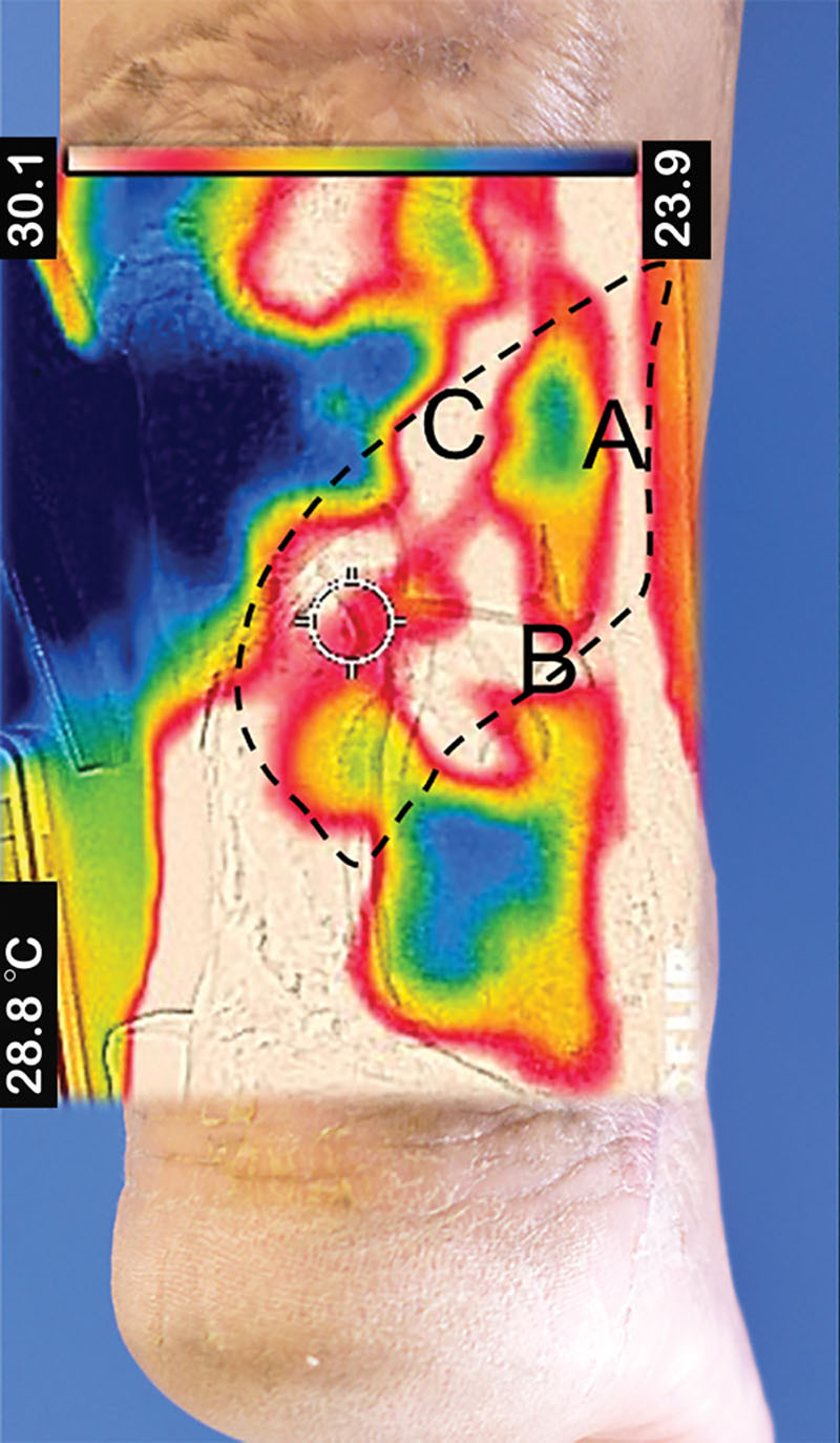
Fig. 1.

Thermal image acquired from posterior aspect of a patient’s right ankle. This patient had a chronic wound from an open fracture distal tibia Gustilo 3B with Achilles tendon rupture, which previously failed primary wound closure and wound dressing for 1 year. We brought the patient for free-style flap coverage. The flap was based on peroneal artery perforator (A), lateral calcaneal artery perforator (B), and lesser saphenous vein (C). The dashed line in the flap territory should include the vascular supply as much as possible.
