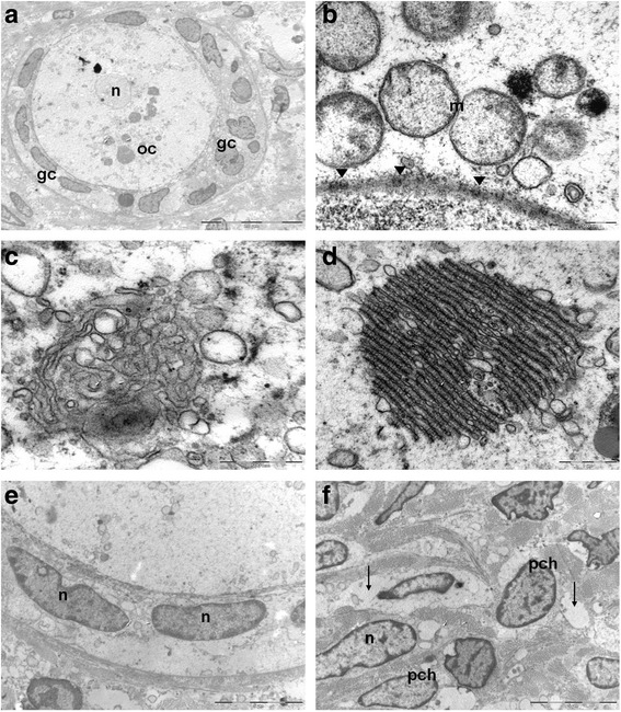
Fig. 4.

Transmission electron microscopy of human cryopreserved ovarian tissue after long-term storage. A primordial/primary follicle: the oocyte shows a nucleus (n) having finely dispersed chromatin and cytoplasm (oc) rich in organelles; a layer of flattened/cuboidal granulosa cells (gc) surrounding oocyte cytoplasm; B perinuclear aggregates of rounded mitochondria (m) having moderate electron dense matrices, ( ) nuclear pores; C Golgi apparatus; D annulate lamellae consist of membrane-bound cisternae traversed by pore complexes; E granulosa cells with oval shaped nuclei (n); f stromal cells with oval shaped nuclei (n) and slight band of peripherically condensed chromatin (pch). Empty interstitial areas are detectable (
 )
)
