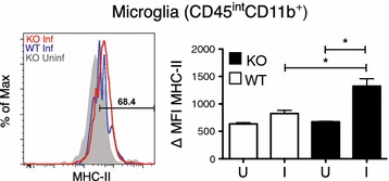
Fig. 3.

Brains from IL-10−/− animals show increased microglia activation during Plasmodium chabaudi infection. Compiled from multiple independent experiments, cells were isolated from PBS-perfused brains of female IL-10−/− (KO) and C57Bl/6J (WT) mice 7–9 days post-infection. Mean fluorescent intensity (MFI) and histogram of MHC-II expression on resident microglia cells (CD11b+CD45int). Histogram colors Uninfected WT (Grey); day 7 and day 9 post-infection WT (Blue); day 7–9 post-infection IL-10−/− (Red). I infected, U uninfected. Student’s t test *p < 0.05, **p < 0.01
