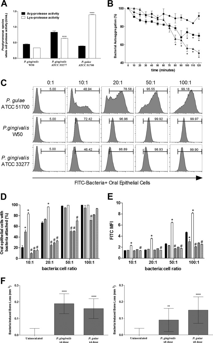
FIG 1.
P. gulae binds to oral epithelial cells, autoaggregates, and has Arg- and Lys-specific proteolytic activity. (A) The proteolytic activity was determined using BApNA (arginine, ■) and LyspNA (lysine, □) substrates. P. gulae whole cells (108 bacteria/ml) showed significantly more lysine activity than P. gingivalis W50 (108 bacteria/ml) and P. gingivalis ATCC 33277 (108 bacteria/ml). All data are expressed as U/ml of proteolytic activity from two separate experiments (n = 6). Means ± the standard errors of the mean (SEM) are shown (****, P < 0.0001). (B) Autoaggregation was measured as the decrease in optical density, with an OD650 set at an initial 1.00 for each bacterium. P. gingivalis W50 (●) displayed minimal autoaggregation, whereas P. gingivalis ATCC 33277 (■) and P. gulae ATCC 51700 (▲) showed significantly more autoaggregation. Data are representative of three separate experiments. Means ± the SEM are shown (*, P < 0.05, P. gulae and P. gingivalis ATCC 33277 versus P. gingivalis W50 [n = 3] and P. gulae and P. gingivalis ATCC 33277 versus P. gingivalis W50 [n = 3]). (C to E) P. gingivalis (W50 and ATCC 33277) and P. gulae ATCC 51700 were labeled with FITC and examined for their ability to bind to oral epithelial cells in increasing ratios of bacteria to epithelial cells. All bacteria were found to bind to oral epithelial cells (C); however, P. gulae ATCC 51700 (□) bound at a greater rate than did P. gingivalis ATCC 33277 () and P. gingivalis W50 (■), as measured by the percentage of FITC-positive cells (D) and the MFI (E). Bacteria treated with TLCK—P. gulae ATCC 51700 (▨), P. gingivalis W50 ( ), and P. gingivalis ATCC 33277 (▩)—showed a decrease in binding. Data are expressed as the percentages of FITC-positive cells or the MFIs and are representative of three separate experiments. Means ± the SEM are shown (*, P < 0.05 [P. gulae and P. gingivalis ATCC 33277 versus P. gingivalis W50]; #, P < 0.05 [TLCK-treated bacteria versus non-TLCK-treated bacteria]; n = 3). (F) BALB/c mice were orally inoculated with either four or eight doses of 1010
P. gulae ATCC 51700 or P. gingivalis W50 cells in two separate experiments. Maxillary bone loss was measured 8 weeks after the first inoculation; data are expressed as bacterium-induced bone loss in mm2. Means ± the SEM are shown (**, P < 0.005; ****, P < 0.00005 [P. gulae and P. gingivalis W50 versus uninoculated; n = 10]).

