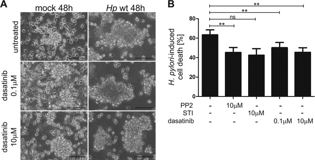
FIG 5.
SFK and c-Abl activities play a crucial role in cell death of H. pylori-infected MEC1 cells. (A) MEC1 cells were treated with 0.1 μM or 10 μM dasatinib prior to infection or left untreated. Phase-contrast microscopy was performed after 48 h of infection. Bar, 100 μm. (B) MEC1 cells were pretreated with 10 μM PP2, 10 μM STI-571, or 0.1 μM or 10 μM dasatinib or remained untreated (−). After infection for 48 h, cell proliferation was measured by an MTT assay. H. pylori-infected cell levels were normalized to those of the respective noninfected controls treated with the same inhibitor. Results represent the means ± standard deviations of three independent experiments performed in quadruplicates. **, P < 0.01; ns, not significant.

