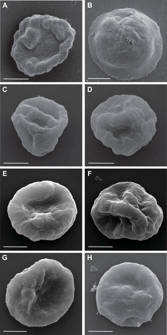
FIG 2.

Surface characteristics of PEs from different P. falciparum parasite lines. Shown are scanning electron micrographs of the P. falciparum 3D7 control (A), P. falciparum NF54 (cultured cell bank) (B), P. falciparum NF54-S01 (derived ex vivo from S01 at the time of drug treatment) (C), P. falciparum NF54-S02 (derived ex vivo from S02 at the time of drug treatment) (D), P. falciparum 3D7B (cultured cell bank) (E), P. falciparum 3D7-S102 (derived ex vivo from S102 at the time of drug treatment) (F), P. falciparum 7G8 (cultured cell bank) (G), and P. falciparum HMP02 (ex vivo cell bank) (H). Representative images are shown, and the scale bars all represent 2 μm.
